Comments on a paper - Macrodontia in guinea pigs (Cavia porcellus) – Radiological findings and localization ion 131 patients. (Journal of Exotic Pet Medicine 39 (2021) 68-75).
[In der Blog-Übersicht wird hier ein Weiterlesen-Link angezeigt]
Full article at Journal of Exotic Pet Medicine
Abstract:
Background: Guinea pigs with dental disease usually present with nonspecific signs such as reduced food intake, difficulty eating and poor general condition. It is assumed that deficient dental attrition is the cause for overlong teeth. Giant teeth, or Macrodonts, have been mentioned as an occasionally occurring pathology.
Methods: Guinea pigs with clinically and radiologically confirmed dental disease presented between 2011 and 2014 and with complete sets of radiographs of the skull (laterolateral, dorsoventral, oblique and isolated views of the mandible and maxilla) were included in this retrospective study (n = 131). Every tooth was radiologically assessed and documented as changed, if an alteration in tooth width, tooth structure, periodontal ligament space, alveolar bone lamina dura and, in the incisors, pulp cavity was present. Special focus was placed on macrodonts and their additional alterations besides increase in width.
Results: The majority (89% = 116/131) of all guinea pigs had at least one macrodont; more than half (58% = 246/422) of all abnormal teeth were macrodonts. In total, 246 teeth (54 incisors, 192 cheek teeth) were diagnosed as macrodonts by means of radiography based on the presence of tooth expansion. Mandibular incisors were much more often affected by macrodontia (87%, 47/54). Furthermore, 91% (175/192) of cheek teeth macrodonts were found in the last two tooth positions. In the present study 97% of all macrodonts (238/246) showed both expansion and structural loss. Alterations to the periodontal ligament space were found in 76% (118/246) of macrodonts. The lamina dura of alveolar bone was changed in 46% (113/246) of cases. Abnormalities of the pulp cavity were present in 80% (43/54) of incisor macrodonts.
Conclusion and clinical relevance: In conclusion, macrodonts occur much more often than previously documented and present a common diagnostic finding in guinea pigs with dental disease. Macrodontia is radiologically characterised by expansion of the tooth and structural loss.
Here some critical comments on this published case:
“Macrodontia in guinea pigs is mentioned only rarely. Extensive literature review yielded only one peer reviewed publication that involves macrodontia in guinea pigs. The following databases ……(11 different)…………. were searched with the keywords: guinea pig, dental disease, radiography, macrodontia and giant tooth and/or teeth on 07.07.2021”. The problem with finding “macrodonty/macrodontia in guinea pigs” in literature is that this term is only a rough clinical / macroscopic “description” of the affected tooth. It just says that the tooth is “enlarged”. This is a term that is too general, non-scientific, in principle. Someone started to use it (who? Schweda?) and now it is more or less generally used to denote a kind of “diagnosis” which is not correct in my opinion. It is like saying that an animal suffers from “long teeth” and search this term in literature. I guess, it is not so simple to say that “macrodontia” is mentioned rarely in literature since it is the most common problem in all guinea pigs suffering from advanced malocclusion. I would say, that it is just difficult to find the individual cases since various descriptions are used. So in my estimation, the keywords used for the search were not appropriate – selected too simply. The authors of this paper themselves tend to use different descriptions for “macrodontia”: extended tooth, expansion, teeth expanded in width, thickening of the infected tooth, massively expanded etc. Other possible options: big tooth, taurodontism, enlarged dental body, Gigantismus dentarius, partial macrodontia, large tooth, dental gigantism etc.
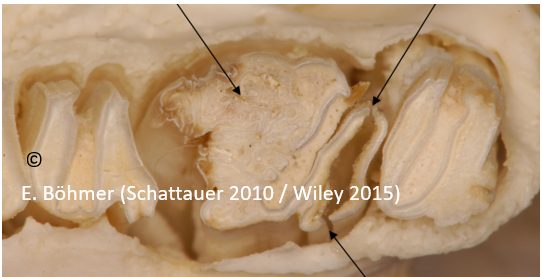
Another example: I described such a “giant tooth” in my dentistry book (Schattauer 2010 and Wiley 2015) (fig. 10.34) as a cheek tooth with a “thickening of the rostral dentin column and longitudinal splitting of the distal tooth body” (fig. 10.34). The same pathology is named “macrodontia” in a paper that tries to emphasize the importance of CT studies (see the CT-image – Schweda et al 2014).
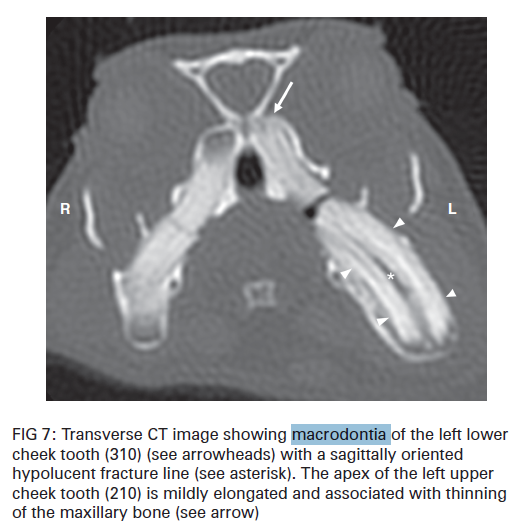
(Schweda et al 2014)
Let us see what “macrodontia” really means: Macrodontia is a situation where a tooth is abnormally enlarged. It can affect single teeth or it can be generalized (partial macrodontia vs. generalized macrodontia). It is a rare inherited condition in humans and seems common with pituitary gigantism, or excess growth hormones and insulin-resistant diabetes among many others. The term is used to designate teeth that are larger than normal (Agurto et al 2019). It is of unknown etiology, but it is also associated with an autosomal dominant pattern of inheritance (Agurto et al 2019). So it seems to be primarily an anomaly or alteration of the tooth size, a genetic mutation. Predominantly affected are permanent teeth which get larger during the tooth development / growth. This hereditary component however, does not (!) apply to guinea pigs. Here we just describe the progressively occouring enlarged size of a single tooth. A tooth that was initially normal and healthy. So far I’ve never seen or diagnosed a macrodont tooth in younger animals with a normocclusion. That is why in my opinion the term “macrodontia” is an absolutely inappropriate denotation for such secondarily “thickened” teeth. Especially, as sometimes teeth are altered structurally in the same way, but remain normal in size or get even smaller / thinner. If we concentrate only on “macrodonts” we will miss these pathologies.
“The authors propose to define macrodonts as teeth with expansion on width and structural deficiency on radiographic examination”. This can be proposed and done, but it is not helpful for a detailed assessment of malocclusions in guinea pigs. People have teeth with dimensions which are proportional to the size of their jaws. This helps to identify enlarged teeth. But what is the exact definition of “enlarged teeth” in guinea pigs? Just the comparison with their neighbors? and the existence of additional changes in structure? See here an example (Fig 5 of the paper):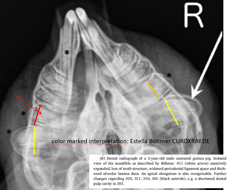
I guess, we all agree that both last molars are “macrodont” teeth (yellow line). I wonder why the authors marked only the right molar with a white arrow? Has the also altered left "macrodont" molar not been included in the statistics? Maybe we all agree that the last but one right molar (M2) is “macrodont” as well. But how would you assess the last but one left cheek tooth? Is it a “macrodont”, too? or a healthy molar? (red line). It shows definitely changes in its structure (dotted red arrow) why we can state in any case that this tooth is abnormal and absolutely not healthy. However, this tooth is not really enlarged. Is it part of the statistic? And what about the right premolar (P4)? It is structurally changed (red arrow) and enlarged (red dotted line) in comparison to the nearby first molar (white dotted line). A macrodont as well?
It is very important to notice that this radiograph depicts different stages of one and the same pathology. All five cheek teeth are affected, but only two appear really enlarged (“macrodonts”). Does it make sense to mention only those two macroscopically altered cheek teeth as “macrodonts” and to sweep the other three also altered cheek teeth under the mat/carpet? Only because they show an earlier stage of disease? This is not reasonable and helpful, in my opinion. Therefore the significance of the statistics of this paper must be viewed very critically. One could compare it with a statistic about tartar where only teeth with massive tartar deposits are recorded. This doesn’t make sense.
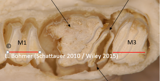
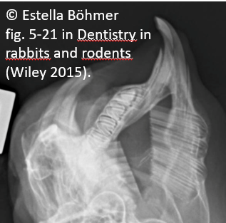
Let us go back to the specimen of a guinea pig with a far advanced alteration of both caudal cheek teeth. The last but one molar (M2) is a “macrodont” based on the description in this paper. And what about the last ipsilateral cheek tooth (M3)? Is it a healthy tooth? Definitely not! It is a highly altered cheek tooth showing an advanced stage of structural change. But based on the definition of “macrodontia” in this paper, this tooth would definitely not be recognized as abnormal since its width is even smaller in comparison to that of the other healthy teeth. Just compare its size with the first molar (M1). The last molar (M3) is definitely smaller (both red lines). Should we name this “microdontia” now? It is the same nonsense as the description of a “macrodontia”. These are both teeth that are the result (!) of far advanced malocclusion with an increased axial load on their dental bodies and a secondary irritation of the germinal tissue. And if we make a statistic about these findings, we should focus more on the structural change of the teeth, regardless of their size. This would help to detect all altered teeth within a dentition and only this would make sense for me. Based on this, the statistics of this paper are basically useless (in my opinion). They show only the final stage and ignore all other stages before this. They only show teeth “with massive tartar” (as mentioned above).
In my opinion, the definition “compound or complex odontoma” (depending on the individual situation) seems more appropriate than “macrodontia”. Compound odontoma is a regularly calcified tissue that bears similarity to teeth. It is a tooth-like structure with a more or less physiological arrangement. Odontomas seem to be developmental anomalies (hamartomas) and not true neoplasms. They are composed of an overgrowth of mature tissue with disorganization and often one element predominating (Anreoli et al 2000). Based on the degree of morphodifferentiation or on the basis of their resemblance to normal teeth, they are divided into compound and complex odontomas (Khanum et al .2014).
The exact mechanism that causes odontoma formation is still unknown, but trauma during tooth development (primary dentition) is a possible causative factor. This fits in with the fact that macrodont teeth in guinea pigs can be mostly found where they are exposed to particularly high pressure which causes persistent damage to the germinal tissue. Also in this study 91% of the two last cheek teeth showed signs of “macrodontia” due to assumed increased compressive stress.
“The identified abnormal findings were scored as either present or absent and were not scored for severity. Width of single teeth was assessed by means of comparison with the other cheek teeth. Since there are no reference measurements for guinea pig tooth width, the evaluation was subjective. The occurrence of a finding was considered as present as soon as it was clearly detected in one of the radiographs”. That is very interesting, but not a very good approach to recognize all “macrodont” teeth (in my opinion). Based on this I have to doubt the results of this analysis (see the discussion above). See for explanation image 2 B of this paper:
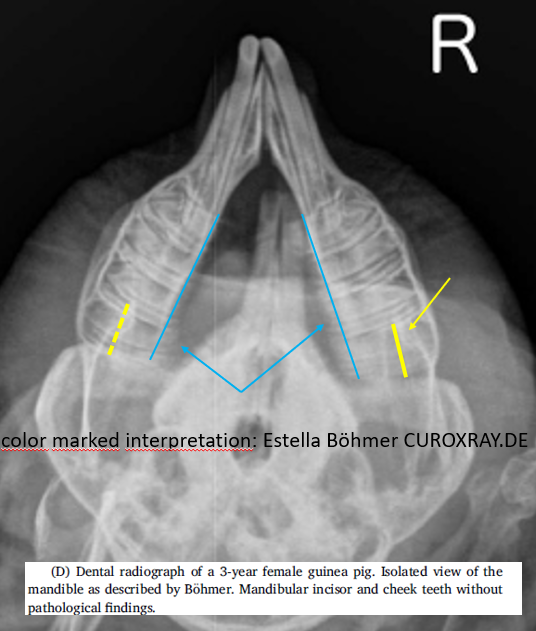
Caption says: “Mandibular incisor and cheek teeth without pathological findings!” Really??? Do you agree? Have a look at the width of both last molars (M3) (yellow lines). They differ in size. The right last molar (M3) is definitely “enlarged” and therefore should be named “macrodont”, already because of its additional structural change (yellow arrow). Unfortunately, this tooth, however, is not part of the statistics since it is assessed as “normal”. In addition to that, all cheek teeth on the right and the last cheek tooth on the left show intraoral tooth elongation (blue lines and arrows). This is a misinterpretation! And based on this the statistics in this paper must be questioned.
It is also important to note that in order to assess the cheek teeth in more detail, the isolated view of the mandible according to Böhmer can be taken additionally by rotating the head about 40 degrees to the right and left, respectively. This allows a much better assessment of single suspect teeth. I miss a reference to this special positioning in the paper. Was a corresponding oblique isolated view of the mandible according to Böhmer also taken in all patients of this study, which is mandatory for a more precise diagnosis? This is part of a normal diagnostic process in guinea pigs with malocclusion visible on standard skull rads in order to focus on a more detailed visualization of all mandibular cheek teeth and incisors.
“In 8 teeth out of 246 identified as macrodonts, expansion of the tooth was the only finding”. I’m pretty sure that structural changes of these teeth have been overlooked (what the authors themselves also cite). So it might have been interesting to include one radiograph of such a finding since it is very unlikely. I agree that “macrodont” teeth always show a structural change. I’ve never diagnosed or read about enlarged healthy teeth in guinea pigs.
“In 97% of the cases, macrodonts showed additional structural changes”. Based on my experience, 100% of all macrodonts show additional structural changes.
“Macrodonts should be considered as cause for dental overgrowth and malocclusion”. I disagree with this statement. “Macrodonts” are the late result of intraoral dental overgrowth since they are the consequence of a highly increased axial pressure on the teeth and their germinal tissue.
“The dental diseased animals found in this study were middle-aged with a mean age of 46 months”. Most guinea pigs suffering from “recognizable/visible” malocclusion are about 3-4 years of age (Böhmer at al. 1988). This matches with this statement. But this paper on “macrodontia” in guinea pigs lacks statistics on the exact age of the animals showing this kind of “tooth enlargement”. That is a pitty. Because it would certainly show that none of the patients is very young. Because “macrodontia” is a progressively acquired and not an inherited pathology. The teeth in all affected 131 guinea pigs were initially healthy and normal. It is uncomprehensible for me why this important statistic on the age of the patients is missing.
“However, a convenient, explicit, and precise definition of macrodontia is missing so far”. In my opinion, a precise definition is still missing. This paper does not give a helpful answer to this question.
Estella Böhmer